
To understand how the brain turns negative signals into adaptive responses.
- ExteroceptionAversive sensory & social stimuliPain, threat and social stress from the external worldLearn more ↓
- InteroceptionDisrupted internal homeostasisSignals such as thirst from inside the bodyLearn more ↓
- Neurons + astrocytesNeuron–glia interactionsHow astrocytes shape the circuits that respond to these signalsLearn more ↓
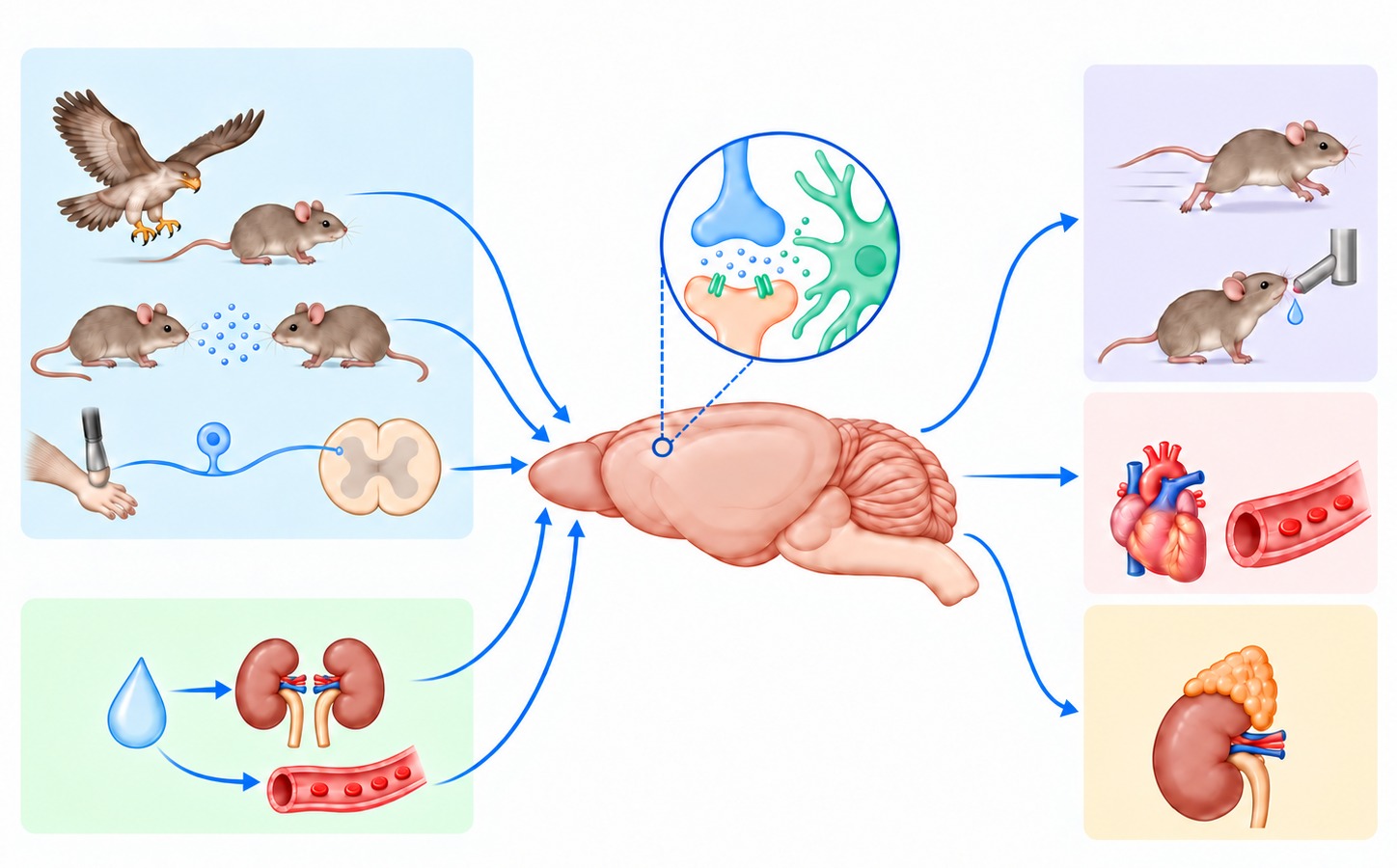 Exteroception
Interoception
Neurons + astrocytes
Behavior
Autonomic response
Hormone release
Illustration created with ChatGPT (OpenAI).
Aversive sensory & social stimuli
Pain and aversive somatosensation
We investigate how noxious and aversive somatic stimuli are detected by peripheral sensory neurons and transmitted through the dorsal root ganglia and spinal cord to higher brain regions.
Our work focuses on the molecular mechanisms that regulate sensory-neuron excitability and on neural circuits that process mechanical sensation, pain, and pathological hypersensitivity.
Aversive visual and social stimuli
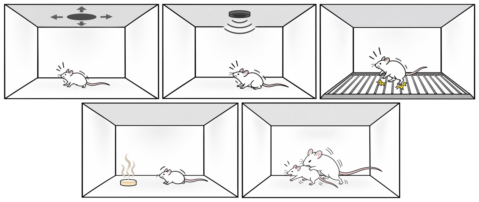
We also study how threatening visual cues and aversive social experiences, such as social stress, are represented by defined neural circuits and transformed into defensive and adaptive responses.
By manipulating and monitoring genetically defined neuronal populations, we seek to understand how sensory information influences affective states, social behavior, and action selection.
Selected publications
- Yang SY, Yun T, Lee Y, … Kim YS*, Park J-Y*, Lee H* (2026). TWIK-1 plays distinct roles in spinal and peripheral sensory circuits controlling mechanical sensitivity and neuropathic hypersensitivity. Signal Transduct Target Ther 11, 248.
- Seo K, Won S, Lee H-Y, … Choi S-Y*, Lee H* (2024). Astrocytic inhibition of lateral septal neurons promotes diverse stress responses. Nat Commun 15, 10091.
- Lee JS, Han JS, Lee K, Bang J, Lee H (2016). The peripheral and central mechanisms underlying itch. BMB Rep 49, 474–487.
- Lee H, Kim D-W, Remedios R, … Anderson DJ (2014). Scalable control of mounting and attack by Esr1+ neurons in the ventromedial hypothalamus. Nature 509, 627–632.
- Cavanaugh DJ†, Lee H†, Lo L†, Shields SD†, … Anderson DJ (2009). Distinct subsets of unmyelinated primary sensory fibers mediate behavioral responses to noxious thermal and mechanical stimuli. PNAS 106, 9075–9080.
Disrupted internal homeostasis
The nervous system continuously monitors the physiological state of the body. When homeostasis is disrupted, it generates coordinated neural, endocrine, autonomic, and behavioral responses to restore balance.
We investigate how signals of homeostatic imbalance are detected and represented by neural circuits, and how these representations drive restorative physiological and behavioral responses.
Thirst: a signal of disrupted fluid homeostasis
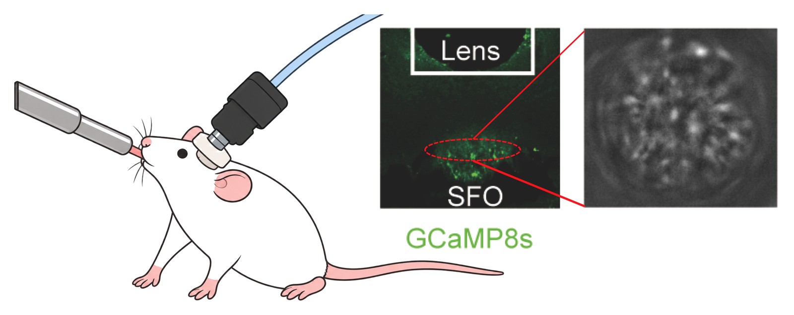
Using thirst as our model, we study how loss of body-fluid balance is detected by the nervous system and translated into compensatory responses such as thirst and drinking behavior.
This system provides a powerful framework for investigating how internal physiological signals are sensed, integrated, and transformed into behavior.
Neuron–glia interactions
Beyond neuron-centric models of neural circuit function
Neural circuits are composed of both neurons and glial cells. We investigate how these cell types interact to shape neural activity, physiological responses, and behavior.
Rather than viewing glial cells as passive support cells, we study how astrocytes sense changes in their local environment, communicate with neighboring neurons, and influence circuit dynamics.
Communication between neurons and glia may matter beyond normal brain function. Growing evidence indicates that neural activity can promote the progression of glioma, a brain tumor arising from glial-lineage cells, opening new questions at the interface of neuroscience and oncology.
Astrocytes in stress-related circuits
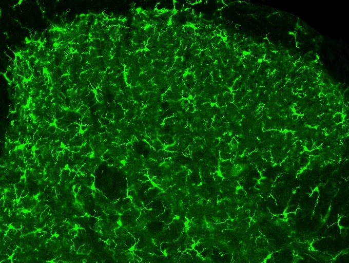
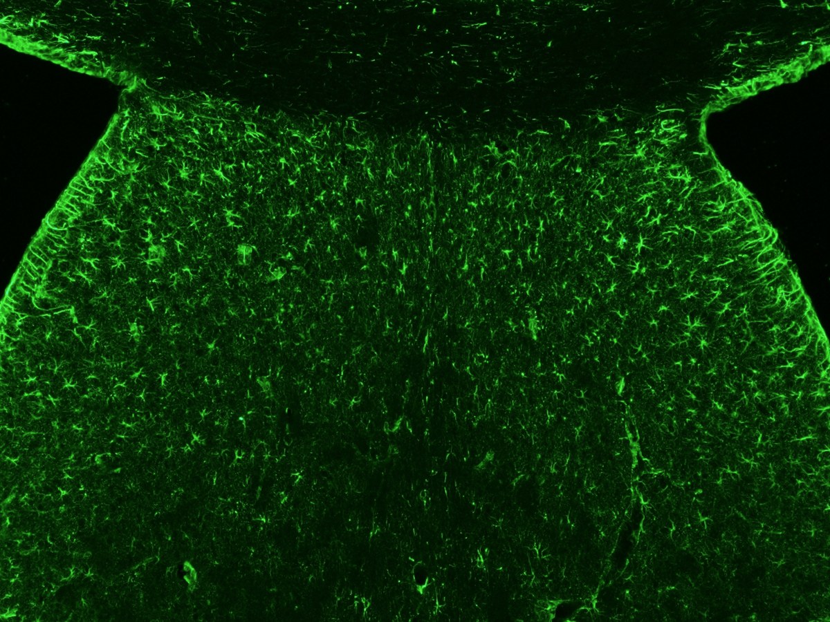
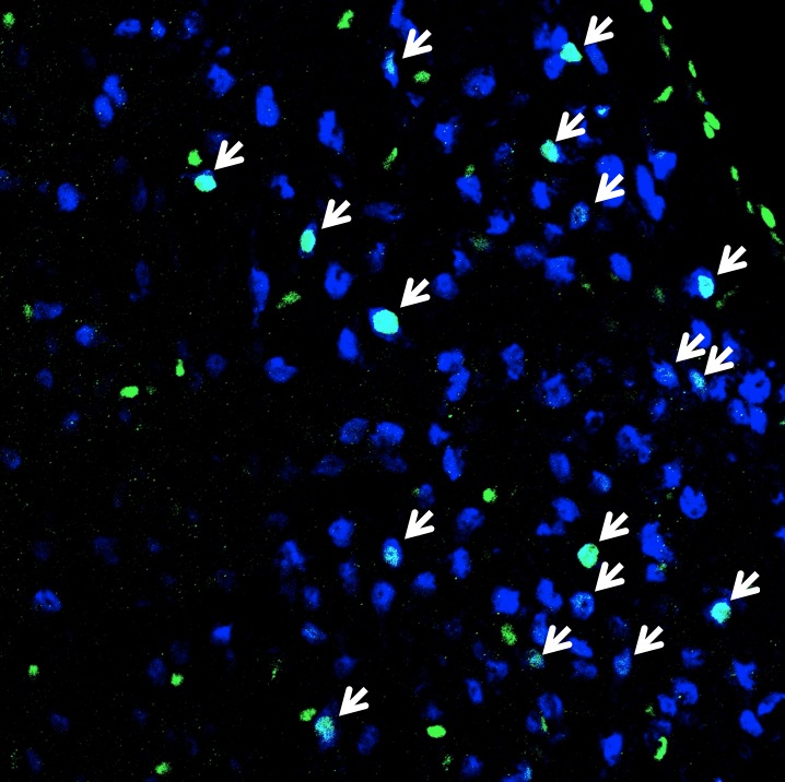
Our work has shown that astrocytes in the lateral septum respond to aversive and social stimuli and regulate neighboring neuronal circuits involved in physiological and behavioral stress responses.
We are extending these studies to understand how neuron–glia interactions contribute to both exteroceptive and interoceptive processing.
Selected publications
- Seo K, Won S, Lee H-Y, … Choi S-Y*, Lee H* (2024). Astrocytic inhibition of lateral septal neurons promotes diverse stress responses. Nat Commun 15, 10091.
- Hwang SN, Lee JS, Seo K, Lee H (2021). Astrocytic regulation of neural circuits underlying behaviors. Cells 10, 296.
- Bang J, Kim HY, Lee H (2016). Optogenetic and chemogenetic approaches for studying astrocytes and gliotransmitters. Exp Neurobiol 25, 205–221.
Experimental approaches
We use the mouse as our primary experimental model and combine molecular, genetic, physiological, imaging, and behavioral approaches to investigate nervous-system function across multiple levels of organization.

Molecular and genetic manipulation
of defined neuronal and glial populations
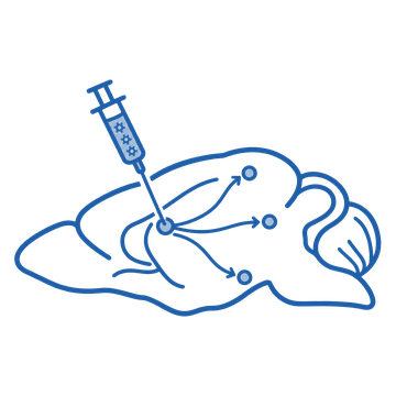
Delivery of viral vectors
to express exogenous proteins and trace neural circuits
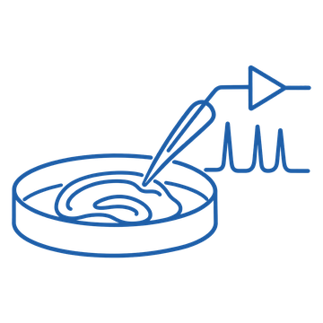
Ex vivo electrophysiology
in acute brain, spinal-cord, and sensory-neuron preparations
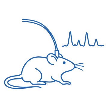
In vivo fiber photometry
to monitor population-level neural and glial activity in behaving animals
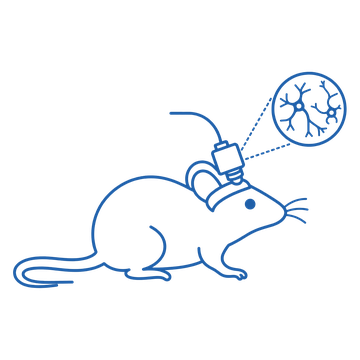
Miniscope calcium imaging
to examine cellular activity and population dynamics during behavior
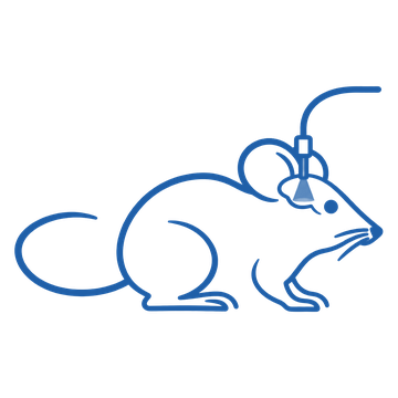
Optogenetic and chemogenetic manipulations
of neural circuits and cell populations
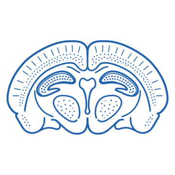
Anatomical and histological analyses
of neural circuits and cell populations

Behavioral assays
for somatosensation, pain, stress, social behavior, thirst, and homeostatic regulation
Icons created with ChatGPT (OpenAI).
By integrating these approaches, we seek to connect molecular and cellular mechanisms with neural-circuit dynamics and ultimately with physiological and behavioral function.